

Surface-enhanced Raman spectroscopy
Surface-enhanced Raman spectroscopy (SERS) is a spectroscopic technique which utilizes the optical properties of metallic surfaces to substantially enhance the Raman signal of species adsorbed on such surfaces. SERS is, in fact, an extension of the classical Raman spectroscopy in which structural information of the target sample is obtained by observing the inelastic light scattering of the incident laser light on the sample’s characteristic vibrational modes (phonons). The Raman effect is intrinsically weak due to the very small cross-section of the inelastic scattering event and therefore does not allow investigation of substances in low concentrations. In SERS, the high intensity of localized surface plasmon electric fields at the surface of metallic nanostructures is used to enhance the Raman signal of the analyte adsorbed on or near the surface. The surface-enhanced signal can be higher than the bare Raman signal by more than ten orders of magnitude, allowing ultrasensitive Raman spectroscopy, going down even to a single molecule. Given that SERS takes place near the surface of metallic nanostructures, the lateral resolution of this technique is determined by the confinement of the localized surface plasmon fields, which are well below the diffraction limit. High sensitivity and the possibility to measure Raman spectra from extremely small volumes make this technique very promising for variety of applications such as nano and bio-sensing and DNA sequencing.
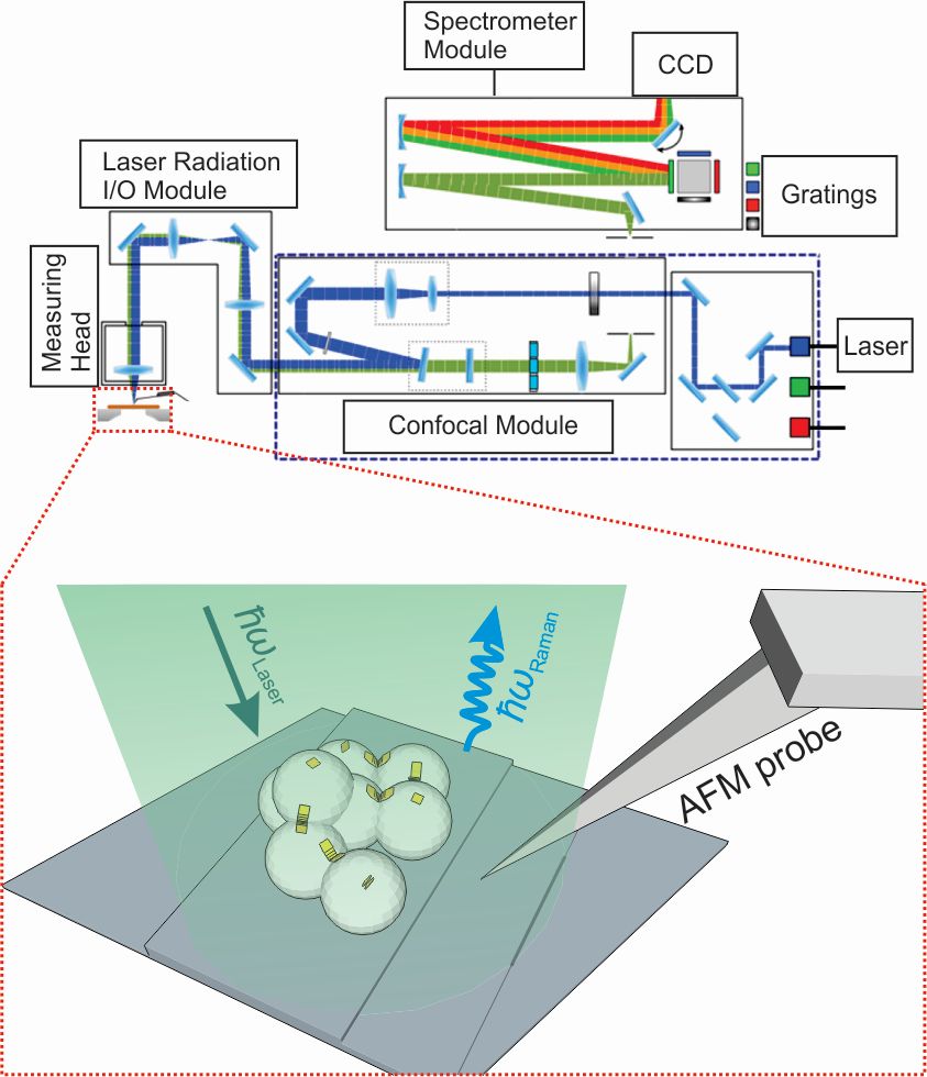
Figure 1. Schematic of the combined SERS-AFM experiment carried out using our NTEGRA Spectra system.
We have, for example, studied dye-metallic nanoparticle complexes using Raman spectroscopy in combination with atomic force microscopy. This is done in collaboration with the Vinča Institute of Nuclear Sciences (http://www.vin.bg.ac.rs/index.php/en/) and within the framework of COST Action MP1302 "NanoSpectroscopy" (http://www.cost-nanospectroscopy.eu). Our focus was on J-aggregation of thiacyanine dyes on the surface of silver citrate capped nanoparticles. Such dye-nanoparticle complexes can have applications ranging from nanoscale sensing to advanced composite materials for novel active and nonlinear optical devices.
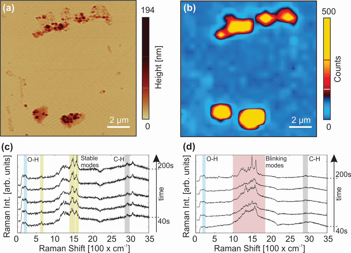
Figure 2. (a) AFM topography image showing several nanoparticle-J-aggregate assemblies on MICA surface, (b) corresponding map of the total Raman intensity showing positions of the “hot spots”, (c) time-resolved SERS spectra acquired for 40 s at one of the “hot spots” in (b), (d) time-resolved SERS spectra of citrate ions. All shown spectra were taken under the same conditions using 532 nm laser with power density of ~200μW/μm2. The spectral signature of citrate ions is identified by (i) the O-H band around 220 cm-1, (ii) the C-H band around 2960 cm-1 and (iii) pronounced blinking in the 1000-1800 cm-1 range. In contrast, dye molecules adsorbed on the nanoparticles are recognized by several stable Raman bands between 200 and 1600 cm-1.
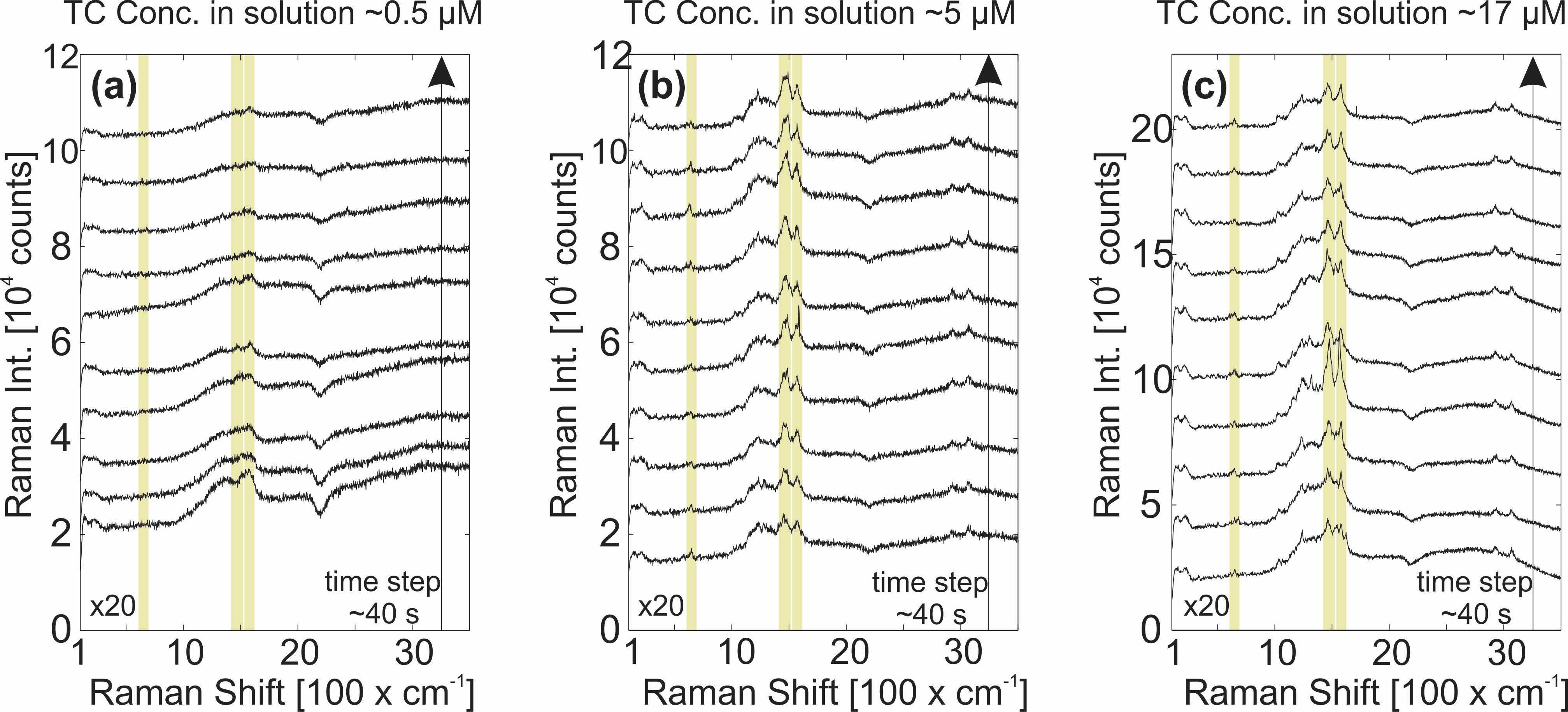
Figure 3. Time resolved SERS spectra (with 40 s time step) at “hot spots” on the samples made by varying the TC concentration in TC-AgNP mixture: (a) ~0.5 μM, (b) ~5 μM and (c) ~17 μM. Highlighted by yellow markers are the characteristic bands of TC J-aggregates. MICA is used as a substrate. All shown spectra were taken under the same conditions using 532 nm laser with power density of ~200μW/μm2.
References:
Hybrid Structures for Surface-Enhanced Raman Scattering: DNA Origami/Gold Nanoparticle Dimer/Graphene
Prinz, J., Matković, A., Pešić, J., Gajić, R. and Bald, I,
Small 12 (39), 5458-5467, (2016) doi:10.1002/smll.201601908