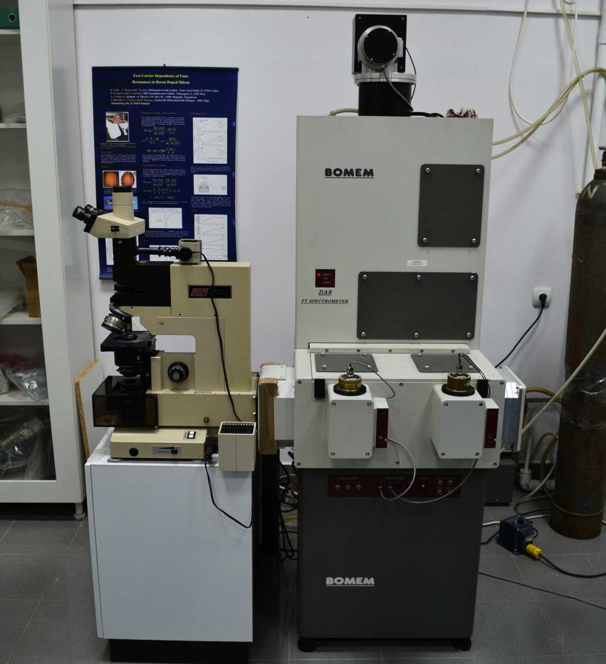


The Bomem DA8 is a research grade Fourier-transform spectrometer for the range 4 to 50 000 cm-1. The term ‘research grade’ refers to an instrument operating under the vacuum, having high resolution, high scanning stability and an access to several input-output ports for several different experiments. It has a vertical conventional Michelson interferometer with a patented dynamical alignment system keeping the exact alignment of the mirrors during each scan. The average angular deviations from an optimal alignment are less than 10-6 radians in normal laboratory environments and about 10-5 radians under severe vibration conditions. A special advantage of the instrument is a unique far infrared Hypersplitter that covers a broad spectral range from 40 to 700 cm-1.
It consists of three sections: the upper one containing the source and the beamsplitter compartment, the middle section with the beam switching compartment, the sample compartment and the detector modules, and the lower part containing the vacuum leads, the power supplies and the data processing and control electronics. Depending of the moving mirror travel, the resolution ranges from 0.06 to 0.0026 cm-1. The instrument has two focused output beams in a sample compartment and three parallel output beams
In the present configuration our DA8 system operates within the 10 to 25 000 cm-1 range with a maximal resolution of 0.02 cm-1. As sources we use a Hg lamp, a Globar (SiC) and a Quartz lamp. The beamsplitters are a 25 μm mylar film (10-125 cm-1), a Hypersplitter (40-700 cm-1) KBr (500-5000 cm-1) and a Quartz (4000 – 25 000 cm-1). The following detectors are used: DTGS (far IR), MCT (77K, mid infrared), InSb (77K, near infrared) and Si photodiode (visible). The spectrometer is equipped with two cryostats: a He flow Janis STDA 100 (LN2 and a LHe, 4-400 K) and an ARS DMX 20 closed-cycle low vibrational cryostat (4.5-300K) with vibration amplitudes in the nanometer range. The system was recently upgraded from far IR and MID IR to the NIR and VIS ranges, namely, a NIR LN2 InSb detector (IPH5000L) for the range 1800-8500 cm-1, a visible Si (IPH5600L) detector for the range 8500-50 000 cm-1 and a Quartz visible beamsplitter (IMB2100L) for the range 4000-25 000 cm-1. For data acquisition and processing, the DA 8 uses the latest working version of the Bomem GRAMS/AI 7.0 software. In addition, a suitable IR-PLAN advance analytical microscope for measuring samples down to 7 µm in size is used.
IR-PLAN analytical microscope
IR-PLAN analytical microscope is a visible light microscope equipped with a high performance infrared sampling accessory designed for operation with FT-IR spectrometer. The microscope performance is directly related to the detection system because usually the analyzing samples are small so it is necessary to use MCT (mercury cadmium telluride) detector. IR-PLAN enables viewing of the exact sample area that will be analyzed and offers the best resolution available in FT-IR microscopy in order to obtain the highest possible quality spectra with the least stray light. IR-PLAN analytical microscope can operate in transmission and reflectance mode.
Instrument specifications
Viewing optics: Standard 15X Reflachromat IR/VIS objective of the Cassegrainian type design for 150X viewing and 10X D-plan achromat glass objective for 100X viewing and visual identification of the sample area with a larger field of view.
Viewing attachments: Binocular viewer has paired 10X eyepieces with crosshair measuring reticle. In combination with the standard 15X objective, provides over 150X visual magnification.
Detection: use of the spectrometer’s detector optics and detector or a dedicated MCT detector.
Illumination: High intensity reflected and transmitted light illumination with variable light intensity and field and aperture stops.
Objective positioning: 4 position rotatable nosepiece.
Sample positioning: Standard 2"x3" travel rectangular rotatable manual stage with stage clip.
Sample masking: Two circular variable apertures for masking capable of being used simultaneously to mask the sample, anywhere in the field of view.
Field of view& sampling area: Nominal 1.3mm field of view with a 15x objective. Sampling area depend on the detector, detector optics and the collecting objective being used.
Purge: the spectrometer’s purge system can be used or own purge system depending on the instrument and coupling.
Sampling mode: Transmission or reflectance.
Microscope support: Rolling work station/table.