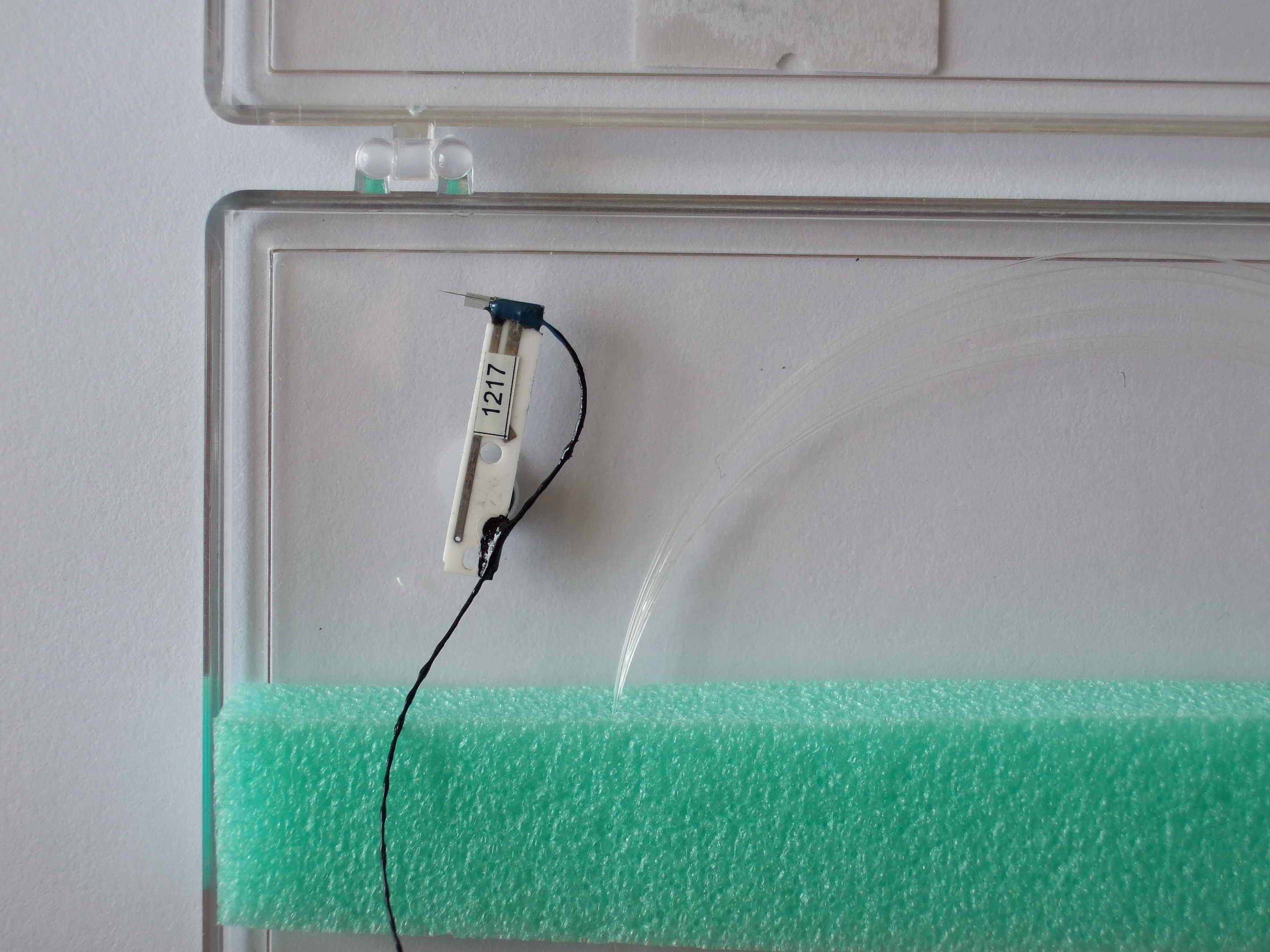


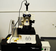
The TwinSNOM system consists of a room-temperature and air-condition Scanning Near-Field Optical Microscope (SNOM) and an Atomic Force Microscope (AFM). The optical microscopy is limited in resolution by the diffraction barrier. The SNOM overcomes this resolution barrier and significantly increases the resolution of the optical microscopy. For a SNOM operation a tapered and metal coated optical fiber is used. The fiber aperture diameter is much smaller than the optical wavelength and this size determines the resolution of the obtained image. In this case, there is no field propagation so the light from the fiber aperture is evanescent and the fiber tip should be brought into the optical near-field of the sample surface. The fiber tip is held in position at the focus of the optics (the reflection objective) for light detection. The sample is then moved with a scanning motion to perform the imaging. A negative feedback loop should be used in order to control the tip to sample distance.
The SNOM fiber tip and the shear-force detector are integrated into a single magnetically mounted and easily exchangeable sensor module. A precise positioning is provided by the use of piezoelectric stepper motors. These motors are used for the remote controlled positioning of the microscope table as well as for the precise positioning of the sensor unit at the focus of the objective. The scanner unit is integrated into the microscope table.
The SNOM control unit contains the laser and the electronics required for light detection. It includes the signal conditioning for the photomultiplier detector as well as a video input selector for the sensor approach monitoring. A highly efficient light collection is achieved in the reflection mode by a specially designed reflection objective.
The TwinSNOM system is placed on a vibrationless table in order to protect the system from the surrounding mechanical vibrations.
The needle sensor completes the functionality of the TwinSNOM. The needle sensor allows for a high resolution non-contact mode atomic force microscopy to be used.
Technical data
Sample scan: lateral: 100 x 100 μm2, vertical: 20 μm, capacitive x/y/z linearisation, 1 nm resolution.
Sample positioning: 30x30 mm2, remote controlled precision piezo drives, step size down to 70 nm.
Tip positioning: remote controlled x/y/z piezo drives, 15x15x10 mm3, step size down to 50 nm.
Aperture diameter: 50 nm nominal.
Shear-force resolution: z: 1 nm, x/y: 10 nm,
Laser: current maximum: 50 mA, max. operating temperature: 50°C, max. radiation flux: ≈1 mW, emission wavelength: 635 nm
Optical microscope: objectives: 50x, 10x (long distance), 10x binocular, conventional mode: upright (bright/dark field), spectral range: 350 nm to 750 nm, detection path: 1700 nm.
Footprint: 55 cm x 55 cm; 60 kg.